IGERT Nanomedicine Trainees and Associates show up big at 2010 NU Research Expo
IGERT Nanomedicine Trainees and Associates show up big at 2010 NU Research Expo
Date: 03/24/2010
Undergraduate Associates Craig Levy and Lauren Moore presenting their work at the 2010 NU Research Expo.
IGERT Nanomedicine Trainees and Associates show up big at 2010 NU Research Expo
Students from the IGERT Nanomedicine program made a big showing at this year’s Northeastern Research Expo. A total of ten PhD Trainees and Undergraduate Associates combined presented their Nanomedicine related research from the Physics, Engineering, Pharmaceutical Sciences and Chemistry and Chemical Biology departments. Please see below for a full listing of student abstracts.
Kathy Chaurasiya (IGERT PhD Trainee)
Nucleic acid chaperone activity of the yeast Ty3 retrotransposon nucleocapsid protein
Reverse transcription in retroviruses and retrotransposons requires nucleic acid chaperones, which facilitate the rearrangement of nucleic acid secondary structure. The nucleic acid chaperone properties of the human immunodeficiency virus type-1 (HIV-1) nucleocapsid protein (NC) have been extensively studied, and nucleic acid aggregation, duplex destabilization, and rapid protein binding kinetics have been identified as major components of its activity. However, the properties of other nucleic acid chaperone proteins, such as retrotransposon Ty3 NC, a likely ancestor of HIV-1 NC, are not well understood. We used single molecule DNA stretching as a method for detailed characterization of Ty3 NC chaperone activity. Wild type Ty3 NC strongly aggregates both double-stranded DNA (dsDNA) and single-stranded DNA (ssDNA), and melted DNA exhibits rapid reannealing in its presence. We also studied several Ty3 NC mutants to identify the roles of functional regions of the protein. The N-terminal basic residues contribute to duplex stabilization, while the zinc finger at the C-terminus counteracts this effect. The mutants examined lack the rapid kinetics of wild type Ty3 NC, indicating that both the basic residues and the zinc finger are required for optimum chaperone activity, which is consistent with previous biochemical experiments. Ty3 NC therefore has a chaperone mechanism similar to that of HIV-1 NC. Although Ty3 NC does not exhibit the strong duplex destabilization of HIV-1 NC, this is consistent with the weaker secondary structure of the Ty3 long-terminal repeat region, which suggests that strong duplex destabilization is not needed for NC to facilitate minus-strand transfer during reverse transcription.
Brendan Harmon (IGERT PhD Trainee)
Correlation of autoradiographic and SPECT imaging of the dopamine transporter in a unilateral 6-hydroxydopamine (6-OHDA) rat model of Parkinson’s disease
The goal of these studies was to assess whether single photon emission computed tomography (SPECT) imaging of the dopamine transporter (DAT) can be used to provide an accurate and sensitivein vivo measure of lesion severity in small animal models of Parkinson’s disease. We adapted our standard protocol for producing 6-OHDA lesions to generate unilateral lesions of the nigrostriatal dopamine neurons with varying degrees of severity. All rats were allowed to recover for three to four weeks post-injection before assessment of right-left differences in striatal DAT radioligand binding using SPECT imaging and autoradiography. For SPECT, rats were given an intravenous injection of 125I- β-CIT, followed by a 2 hour equilibration time. Imaging was performed using a NanoSPECT/CT® scanner (Bioscan, Inc.), and right-left differences in striatal DAT binding were quantified using the instrument’s software. Within 2 hours of completion of the SPECT sessions, the animals were sacrificed and their brains were cut into coronal sections. The sections were placed onto glass slides, dried, and imaged for 1 hour using a Cyclone Phorphorimaging System (Perkin-Elmer). Right-left differences in striatal DAT radioligand binding were quantified from autoradiograms, and the values were compared with those from SPECT analysis of the same brains. A near perfect correlation was achieved (r = 0.989), confirming the ability of the SPECT instrument to accurately measure right-left differences in 125I-β-CIT binding in rat striatum. We conclude that SPECT imaging holds potential as a tool for in vivo assessment of neuroprotective treatments in small animal models of Parkinson’s disease.
Lara Jabr-Milane (IGERT PhD Trainee)
Modulation of Hypoxia-Induced Multi-drug Resistance in Cancer Using EGFR-Targeted Polymer Blend Nanocarriers for Combination Paclitaxel/Lonidamine Therapy
Multidrug resistance (MDR) is most often implicated in cases of recurrent, non-responsive disease and is a significant obstacle in the treatment of cancer. The biological focus of this work is to explore the relationship between the hypoxic microenvironment of a tumor, the development of MDR, and the energetic profile characteristic of the Warburg effect (aerobic glycolysis). The cellular transformation and proteome changes that occur in response to hypoxia were examined. In a panel of nine breast and ovarian cancer cell lines hypoxia was shown to induce MDR and the Warburg effect. Protein analysis of HIF transcriptions factors, MDR markers, EGFR, and glycolytic proteins revealed that the hypoxic transformation was cell-line and duration dependent.
The therapeutic aim of this research is to develop an epidermal growth factor receptor (EGFR)-targeted nanocarrier system for combination (paclitaxel/lonidamine) therapy for the treatment of hypoxic/MDR cancer. EGFR-targeted polymer blend nanoparticles were shown to actively target EGFR expressing cell lines. Targeted nanoparticle uptake emulated the expression of EGFR in the panel of nine cell lines. This nanoparticle targeting was obstructed with competitive EGFR antibody treatment, confirming engagement of the particles with EGFR. Combination therapy with lonidamine and paclitaxel significantly improved the therapeutic index of both drugs.
An orthotopic breast cancer model of hypoxic, MDR cancer was developed in nu/nu mice. This model was confirmed by immunohistochemistry of the tumors for HIF-1α, P-gp, CD-31, EGFR, Hexokinase 2, and GLUT-1. This model is currently being used to conduct a complete pre-clinical evaluation of the drug delivery system.
Joslynn Lee (IGERT PhD Trainee)
Identification of functional subclasses in the ribulose-phosphate binding barrel superfamily using computational tools
The ability to predict the function of a protein from its three-dimensional structure is an important problem in the post-genomic era. This paper describes a computational method for the identification of function for proteins within a superfamily. Superfamilies are defined as a group of evolutionarily related proteins that have similar three-dimensional structures. Although the proteins within a given superfamily all have similar structures, there can be enormous functional diversity within a superfamily. The ribulose-phosphate binding barrel (RPBB) superfamily consists of five subclasses; each of the five subclasses represents a different type of biochemical function. My approach to understanding the functional diversity in this superfamily is based on a local structural analysis at the interaction sites of the individual proteins. First, Theoretical Microscopic Titration Curves (THEMATICS) and Partial Order Optimum Likelihood (POOL) are used to predict the residues involved in catalysis and/or binding for each protein structure in the superfamily. Then, the 3D structures of all of the proteins are aligned. The predicted active residues are labeled on the alignment. From this labeled alignment, patterns emerge that enable the RPBB superfamily to be sorted into functional subclasses. Applications to Structural Genomics (SG) proteins of unknown or uncertain function are reported. For two of the SG proteins in this superfamily, a putative orotidine-monophosphate-decarboxylase and a putative D-ribulose 5-phosphate 3-epimerase, it is shown that the putative functional annotations are likely correct. However, there are several other SG proteins in this superfamily that are shown to be incorrectly annotated.
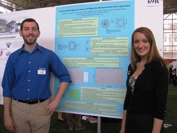
Craig Levy (IGERT Undergraduate Associate)
Electrically triggered release of magnetic Nanoparticles from Liposomes
Magnetic Nanoparticles show promise for application to many areas of medicine, including for use as cancer treatment agents through hyperthermia and targeted drug delivery. When suspended in a fluid these Nanoparticles can be manipulated using externally applied magnetic fields; they can be confined using a static field gradient and can be heated by applying an oscillating field (hyperthermia). It has been shown that these properties apply in vivo, and that they also apply when the particles are contained within certain “nanoplatforms” such as liposomes. Thus, incorporation of magnetic Nanoparticles into liposomes is of interest because it would allow for the creation of a multifunctional nanoplatform which permits magnetic confinement of the liposome and its contents (Nanoparticles, Doxorubicin, etc.) to a desired site (i.e. a tumor site). There is still the problem, however, of how to release the contents of the Magnetic Liposome in a controlled way. Electroporation of vesicles (including liposomes) is a well-studied phenomenon in which application of an external electric field (usually from a short DC pulse) creates reversible pores in the membrane. We have studied the possibility of using AC electroporation to release magnetic Iron (III) Oxide Nanoparticles from liposomes. We see that there is significant release of Nanoparticles from the liposomes and that it is a function of several variables including time, field strength, and degree of field inhomogeneity.
Ragen McAdoo (IGERT Undergraduate Associate)
Gold – Attached Titania Nanotubes for Carbon Monoxide Oxidation
Titania nanotubes have been fabricated by electrochemical annodization in ammonium floride to produce free standing nanotube flakes. Gold nanoparticles 3.5 nm in diameter have been deposited onto the surface of the nanotubes by a deposition-precipitation method engineered in our lab. Gold attached titania nanotubes have been brought to Ron Willey’s lab for Carbon Monoxide oxidation measurements. Measurements were taken at room temperature and have shown up to 75% conversion efficiency.
Lauren Moore (IGERT Undergraduate Associate)
Ligand Exchange Influenced Enhancement of Magnetic Properties in Iron Oxide Nanoparticles for Biomedical Applications.
Magnetic nanoparticles (MagNPs) in the form of iron oxide nanocrystals are used in the next-generation drug delivery systems, MRI contrast agents, and cell separation systems. For the successful implementation of these MagNPs for these applications, it is important that these NPs be synthesized with precise control on their size and size distribution. High-temperature thermal decomposition method in the presence of organic ligands is one approach to synthesis such MagNPs. These MagNPs are soluble in organic solvents and to make them water soluble for biomedical applications a ligand exchange step is necessary. In this presentation, the various surfactants that can be used for ligand exchange and their influence on the magnetic property of these NPs will be presented. Potential mechanism of ligand binding to MagNPs surface and implication of these MagNPs in biomedical applications will be discussed.
Brian Plouffe (IGERT PhD Trainee)
Thermomagnetic Measurement of Magnetic Nanoparticle Size Distribution
Successful employment of nanoparticles in any of their myriad applications (recording media, biomedical processes, catalytic techniques, etc.) is possible only upon accurate determination of the average nanoparticle diameter and its distribution. The most precise, as well as the most time-consuming, determination of these attributes involves study of nanoparticle ensembles using transmission electron microscopy (TEM). Not only does this technique require TEM access, it also requires examination and processing of large amounts of data to properly characterize the ensemble. Previous researchers have determined the average particle size of magnetic nanoparticles from magnetization versus applied magnetic field, M(H). In this work the average particle size and its distribution are determined from magnetization measurements as a function of temperature, M(T). For superparamagnetic nanoparticles the size can be determined from magnetic measurements via the thermal blocking temperature. Employing a model that incorporates the functional distribution of the nanoparticle volume, the average particle diameter and the distribution of diameters of a population of Fe3O4 nanoparticles synthesized by a thermal decomposition method were determined from the magnetic blocking temperature using the zero-field-cooled thermomagnetization data. The results derived from the magnetic measurements compare favorably to those obtained from TEM examination, confirming the value of this approach.
Francisco Reynoso (IGERT PhD Trainee)
Multi-modal MRI, SPECT, CT Imaging of Theranostic Nanoplatforms
The development of non-invasive imaging techniques for the assessment of cancer treatment is rapidly becoming highly important. Magnetic Cationic Liposomes (MCL) are multifunctional and can be used for simultaneous diagnostics (MRI, SPECT/CT), improved delivery (magnetic targeting) and therapy (hyperthermia). Poly-ethyleneglycol (PEG) coated cationic liposomes are loaded with superparamagnetic iron oxide nanoparticles (SPIONS) and tagged with the radioisotope Indium-111. The magnetic properties of the SPIONS make MCL a remarkable MRI contrast agent and useful for magnetically targeting liposomes to specific areas of the body. The radioisotope Indium-111 serves as a contrast agent in SPECT imaging.