Interdisciplinary Collaboration
Our researchers collaborate across five following core disciplines to develop breakthrough imaging tools:
- Probe Development
- Probe Delivery
- Imaging Technologies
- Animal Models
- Signal Processing
THE NEED
Chemical analysis could be used to study processes like neurotransmitter release, but the field currently has a scale problem. New technologies are needed that can balance the specificity of cellular imaging with the functional, structural, or spatial capabilities of whole-body imaging.
CILS Members
Manuscripts enabled by CILS core facilities
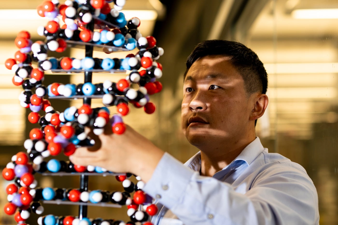
OUR SOLUTION
Our goal is to create a new suite of modular nanoscale tools, probes that image dynamic biological processes beyond what is currently feasible. By forming multidisciplinary teams, we select and focus on a difficult problem that needs a solution – the interface between instrumentation and the biological environment is key to our success. Starting from a research question, we work through the probe’s lifecycle, from novel platform development, continuing through to its biological application.
THE BENEFITS

Enabling a doctor to measure specific biomolecules in a patient over disease progression will lead to better-informed diagnostics and personalized care.

Animal models better reflect a specific neurological disease state, and the same tools used to study these models can be used to further human research.

Diseases can be studied in a highly personalized fashion, for the design and development of precision interventions.
OUR APPROACH
Tissue Clearing Webinar
Come join us on February 10th, 2023 at 1-2 pm (EST)📅 to learn more about how to improve your microscopy images of thick samples via Tissue Clearing 🔬
Our imaging scientist Eun will explain why tissue clearing is important to boost microscopy signal from your thick samples, and will discuss different methods and applications.
Register here: ➡ https://lnkd.in/ey5eg9y2 ⬅

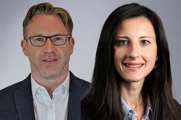
Niedre and Bellini Receive $2.7M Grant from the National Cancer Institute
Mark Niedre
CILS member and bioengineering professor Mark Niedre was awarded a $2.7M grant for 5 years along with co-PI Chiara Bellini. The grant project is titled "Continuous, Non-Invasive Optical Monitoring of Circulating Tumor Cell-Mediated Metastasis in Awake Mice".
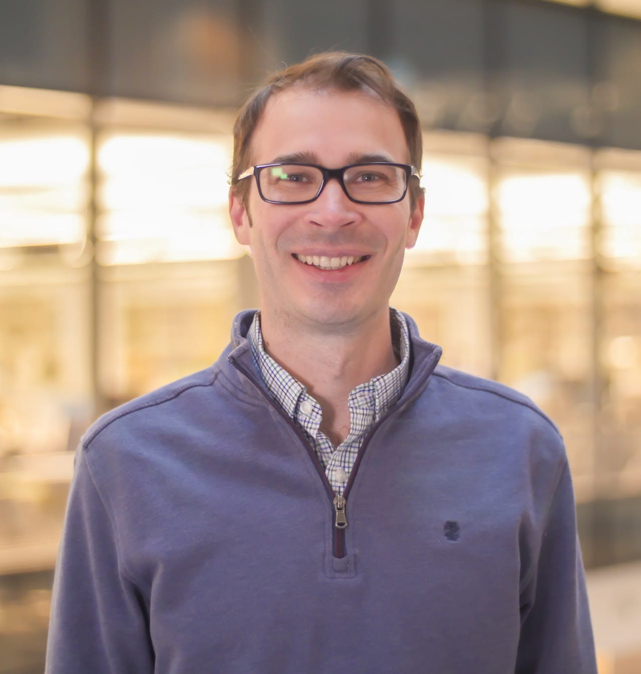
Bryan Spring Named A Scialog: Advancing Bioimaging Fellow
For early-career researchers chosen to address challenges involved in enhancing high-resolution imaging of tissue.
Our facility
Interdisciplinary Science and Engineering Complex
ISEC 080, 805 Columbus Ave, Boston, MA, 02115